| From | xxxxxx <[email protected]> |
| Subject | How Do Human Egg Cells Stay Healthy for Decades? |
| Date | August 2, 2025 12:50 AM |
Links have been removed from this email. Learn more in the FAQ.
Links have been removed from this email. Learn more in the FAQ.
[[link removed]]
HOW DO HUMAN EGG CELLS STAY HEALTHY FOR DECADES?
[[link removed]]
Sahana Sitaraman
July 18, 2025
The Scientist
[[link removed]]
*
[[link removed]]
*
[[link removed]]
*
*
[[link removed]]
_ Contrary to long-standing assumptions, human egg cells stay
pristine for decades not by ramping up waste disposal, but by dialing
it down. _
human egg cell,
A human female is born with all the egg cells she will ever have. The
possibility for the development of new oocytes is zero. Given this
constraint, it is crucial that these gametes remain healthy and viable
for decades until they are needed to form an embryo. Irrespective of
the ‘age’ of the fertilized oocyte, the resulting embryo has the
characteristics of a freshly born cell, indicating the existence of
mechanisms that counteract accrued cellular damage and keep the egg
fresh. What are these processes that drive the prolonged life of human
egg cells?
Elvan Böke
[[link removed]], an
oocyte biologist at the Centre for Genomic Regulation, studies exactly
that. A healthy cell boasts vigilant scanning for and removal of
misfolded, damaged, or unnecessary proteins. A common feature
associated with cellular aging
[[link removed]] is
the breakdown of intracellular protein degradation machinery.1 In
previous studies done in mouse oocytes, Böke and other researchers
found that these cells rely on two key adaptations to keep their
cytoplasm free of harmful clutter: They store and degrade of protein
aggregates in vesicles and contain oocyte proteins with exceptionally
long lives
[[link removed]].2,3
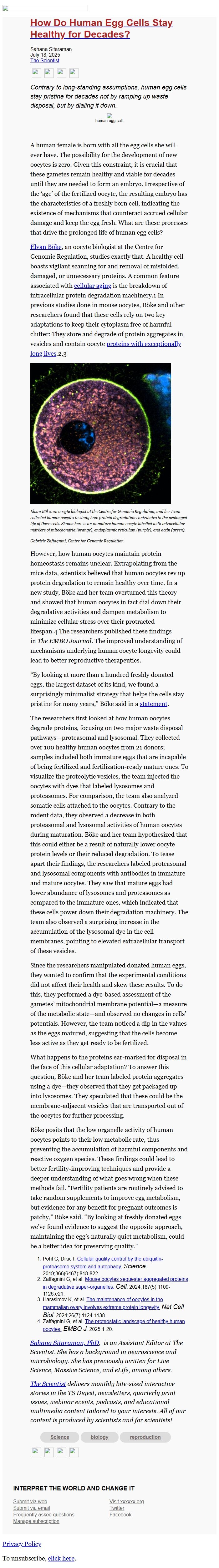
[A microscopic image of an immature human oocyte showing the
localization of diverse intracellular organelles.]
Elvan Böke, an oocyte biologist at the Centre for Genomic Regulation,
and her team collected human oocytes to study how protein degradation
contributes to the prolonged life of these cells. Shown here is an
immature human oocyte labelled with intracellular markers of
mitochondria (orange), endoplasmic reticulum (purple), and actin
(green).
Gabriele Zaffagnini, Centre for Genomic Regulation
However, how human oocytes maintain protein homeostasis remains
unclear. Extrapolating from the mice data, scientists believed that
human oocytes rev up protein degradation to remain healthy over time.
In a new study, Böke and her team overturned this theory and showed
that human oocytes in fact dial down their degradative activities and
dampen metabolism to minimize cellular stress over their protracted
lifespan.4 The researchers published these findings in _The EMBO
Journal_. The improved understanding of mechanisms underlying human
oocyte longevity could lead to better reproductive therapeutics.
“By looking at more than a hundred freshly donated eggs, the largest
dataset of its kind, we found a surprisingly minimalist strategy that
helps the cells stay pristine for many years,” Böke said in
a statement
[[link removed]].
The researchers first looked at how human oocytes degrade proteins,
focusing on two major waste disposal pathways—proteasomal and
lysosomal. They collected over 100 healthy human oocytes from 21
donors; samples included both immature eggs that are incapable of
being fertilized and fertilization-ready mature ones. To visualize the
proteolytic vesicles, the team injected the oocytes with dyes that
labeled lysosomes and proteasomes. For comparison, the team also
analyzed somatic cells attached to the oocytes. Contrary to the rodent
data, they observed a decrease in both proteasomal and lysosomal
activities of human oocytes during maturation. Böke and her team
hypothesized that this could either be a result of naturally lower
oocyte protein levels or their reduced degradation. To tease apart
their findings, the researchers labeled proteasomal and lysosomal
components with antibodies in immature and mature oocytes. They saw
that mature eggs had lower abundance of lysosomes and proteasomes as
compared to the immature ones, which indicated that these cells power
down their degradation machinery. The team also observed a surprising
increase in the accumulation of the lysosomal dye in the cell
membranes, pointing to elevated extracellular transport of these
vesicles.
Since the researchers manipulated donated human eggs, they wanted to
confirm that the experimental conditions did not affect their health
and skew these results. To do this, they performed a dye-based
assessment of the gametes’ mitochondrial membrane potential—a
measure of the metabolic state—and observed no changes in cells’
potentials. However, the team noticed a dip in the values as the eggs
matured, suggesting that the cells become less active as they get
ready to be fertilized.
What happens to the proteins ear-marked for disposal in the face of
this cellular adaptation? To answer this question, Böke and her team
labeled protein aggregates using a dye—they observed that they get
packaged up into lysosomes. They speculated that these could be the
membrane-adjacent vesicles that are transported out of the oocytes for
further processing.
Böke posits that the low organelle activity of human oocytes points
to their low metabolic rate, thus preventing the accumulation of
harmful components and reactive oxygen species. These findings could
lead to better fertility-improving techniques and provide a deeper
understanding of what goes wrong when these methods fail. “Fertility
patients are routinely advised to take random supplements to improve
egg metabolism, but evidence for any benefit for pregnant outcomes is
patchy,” Böke said. “By looking at freshly donated eggs we’ve
found evidence to suggest the opposite approach, maintaining the
egg’s naturally quiet metabolism, could be a better idea for
preserving quality.”
* Pohl C, Dikic I. Cellular quality control by the
ubiquitin-proteasome system and autophagy.
[[link removed]] _Science_.
2019;366(6467):818-822.
* Zaffagnini G, et al. Mouse oocytes sequester aggregated proteins
in degradative super-organelles.
[[link removed](24)00068-0?_returnURL=https%3A%2F%2Flinkinghub.elsevier.com%2Fretrieve%2Fpii%2FS0092867424000680%3Fshowall%3Dtrue] _Cell_.
2024;187(5):1109-1126.e21.
* Harasimov K, et al. The maintenance of oocytes in the mammalian
ovary involves extreme protein longevity.
[[link removed]] _Nat Cell Biol_.
2024;26(7):1124-1138.
* Zaffagnini G, et al. The proteostatic landscape of healthy human
oocytes.
[[link removed]]_ EMBO
J_. 2025:1-20.
_Sahana Sitaraman, PhD
[[link removed]], is an
Assistant Editor at The Scientist. She has a background in
neuroscience and microbiology. She has previously written for Live
Science, Massive Science, and eLife, among others._
_The Scientist [[link removed]] delivers
monthly bite-sized interactive stories in the TS Digest, newsletters,
quarterly print issues, webinar events, podcasts, and educational
multimedia content tailored to your interests. All of our content is
produced by scientists and for scientists! _
* Science
[[link removed]]
* biology
[[link removed]]
* reproduction
[[link removed]]
*
[[link removed]]
*
[[link removed]]
*
*
[[link removed]]
INTERPRET THE WORLD AND CHANGE IT
Submit via web
[[link removed]]
Submit via email
Frequently asked questions
[[link removed]]
Manage subscription
[[link removed]]
Visit xxxxxx.org
[[link removed]]
Twitter [[link removed]]
Facebook [[link removed]]
[link removed]
To unsubscribe, click the following link:
[link removed]
HOW DO HUMAN EGG CELLS STAY HEALTHY FOR DECADES?
[[link removed]]
Sahana Sitaraman
July 18, 2025
The Scientist
[[link removed]]
*
[[link removed]]
*
[[link removed]]
*
*
[[link removed]]
_ Contrary to long-standing assumptions, human egg cells stay
pristine for decades not by ramping up waste disposal, but by dialing
it down. _
human egg cell,
A human female is born with all the egg cells she will ever have. The
possibility for the development of new oocytes is zero. Given this
constraint, it is crucial that these gametes remain healthy and viable
for decades until they are needed to form an embryo. Irrespective of
the ‘age’ of the fertilized oocyte, the resulting embryo has the
characteristics of a freshly born cell, indicating the existence of
mechanisms that counteract accrued cellular damage and keep the egg
fresh. What are these processes that drive the prolonged life of human
egg cells?
Elvan Böke
[[link removed]], an
oocyte biologist at the Centre for Genomic Regulation, studies exactly
that. A healthy cell boasts vigilant scanning for and removal of
misfolded, damaged, or unnecessary proteins. A common feature
associated with cellular aging
[[link removed]] is
the breakdown of intracellular protein degradation machinery.1 In
previous studies done in mouse oocytes, Böke and other researchers
found that these cells rely on two key adaptations to keep their
cytoplasm free of harmful clutter: They store and degrade of protein
aggregates in vesicles and contain oocyte proteins with exceptionally
long lives
[[link removed]].2,3
[A microscopic image of an immature human oocyte showing the
localization of diverse intracellular organelles.]
Elvan Böke, an oocyte biologist at the Centre for Genomic Regulation,
and her team collected human oocytes to study how protein degradation
contributes to the prolonged life of these cells. Shown here is an
immature human oocyte labelled with intracellular markers of
mitochondria (orange), endoplasmic reticulum (purple), and actin
(green).
Gabriele Zaffagnini, Centre for Genomic Regulation
However, how human oocytes maintain protein homeostasis remains
unclear. Extrapolating from the mice data, scientists believed that
human oocytes rev up protein degradation to remain healthy over time.
In a new study, Böke and her team overturned this theory and showed
that human oocytes in fact dial down their degradative activities and
dampen metabolism to minimize cellular stress over their protracted
lifespan.4 The researchers published these findings in _The EMBO
Journal_. The improved understanding of mechanisms underlying human
oocyte longevity could lead to better reproductive therapeutics.
“By looking at more than a hundred freshly donated eggs, the largest
dataset of its kind, we found a surprisingly minimalist strategy that
helps the cells stay pristine for many years,” Böke said in
a statement
[[link removed]].
The researchers first looked at how human oocytes degrade proteins,
focusing on two major waste disposal pathways—proteasomal and
lysosomal. They collected over 100 healthy human oocytes from 21
donors; samples included both immature eggs that are incapable of
being fertilized and fertilization-ready mature ones. To visualize the
proteolytic vesicles, the team injected the oocytes with dyes that
labeled lysosomes and proteasomes. For comparison, the team also
analyzed somatic cells attached to the oocytes. Contrary to the rodent
data, they observed a decrease in both proteasomal and lysosomal
activities of human oocytes during maturation. Böke and her team
hypothesized that this could either be a result of naturally lower
oocyte protein levels or their reduced degradation. To tease apart
their findings, the researchers labeled proteasomal and lysosomal
components with antibodies in immature and mature oocytes. They saw
that mature eggs had lower abundance of lysosomes and proteasomes as
compared to the immature ones, which indicated that these cells power
down their degradation machinery. The team also observed a surprising
increase in the accumulation of the lysosomal dye in the cell
membranes, pointing to elevated extracellular transport of these
vesicles.
Since the researchers manipulated donated human eggs, they wanted to
confirm that the experimental conditions did not affect their health
and skew these results. To do this, they performed a dye-based
assessment of the gametes’ mitochondrial membrane potential—a
measure of the metabolic state—and observed no changes in cells’
potentials. However, the team noticed a dip in the values as the eggs
matured, suggesting that the cells become less active as they get
ready to be fertilized.
What happens to the proteins ear-marked for disposal in the face of
this cellular adaptation? To answer this question, Böke and her team
labeled protein aggregates using a dye—they observed that they get
packaged up into lysosomes. They speculated that these could be the
membrane-adjacent vesicles that are transported out of the oocytes for
further processing.
Böke posits that the low organelle activity of human oocytes points
to their low metabolic rate, thus preventing the accumulation of
harmful components and reactive oxygen species. These findings could
lead to better fertility-improving techniques and provide a deeper
understanding of what goes wrong when these methods fail. “Fertility
patients are routinely advised to take random supplements to improve
egg metabolism, but evidence for any benefit for pregnant outcomes is
patchy,” Böke said. “By looking at freshly donated eggs we’ve
found evidence to suggest the opposite approach, maintaining the
egg’s naturally quiet metabolism, could be a better idea for
preserving quality.”
* Pohl C, Dikic I. Cellular quality control by the
ubiquitin-proteasome system and autophagy.
[[link removed]] _Science_.
2019;366(6467):818-822.
* Zaffagnini G, et al. Mouse oocytes sequester aggregated proteins
in degradative super-organelles.
[[link removed](24)00068-0?_returnURL=https%3A%2F%2Flinkinghub.elsevier.com%2Fretrieve%2Fpii%2FS0092867424000680%3Fshowall%3Dtrue] _Cell_.
2024;187(5):1109-1126.e21.
* Harasimov K, et al. The maintenance of oocytes in the mammalian
ovary involves extreme protein longevity.
[[link removed]] _Nat Cell Biol_.
2024;26(7):1124-1138.
* Zaffagnini G, et al. The proteostatic landscape of healthy human
oocytes.
[[link removed]]_ EMBO
J_. 2025:1-20.
_Sahana Sitaraman, PhD
[[link removed]], is an
Assistant Editor at The Scientist. She has a background in
neuroscience and microbiology. She has previously written for Live
Science, Massive Science, and eLife, among others._
_The Scientist [[link removed]] delivers
monthly bite-sized interactive stories in the TS Digest, newsletters,
quarterly print issues, webinar events, podcasts, and educational
multimedia content tailored to your interests. All of our content is
produced by scientists and for scientists! _
* Science
[[link removed]]
* biology
[[link removed]]
* reproduction
[[link removed]]
*
[[link removed]]
*
[[link removed]]
*
*
[[link removed]]
INTERPRET THE WORLD AND CHANGE IT
Submit via web
[[link removed]]
Submit via email
Frequently asked questions
[[link removed]]
Manage subscription
[[link removed]]
Visit xxxxxx.org
[[link removed]]
Twitter [[link removed]]
Facebook [[link removed]]
[link removed]
To unsubscribe, click the following link:
[link removed]
Message Analysis
- Sender: Portside
- Political Party: n/a
- Country: United States
- State/Locality: n/a
- Office: n/a